Exocrine functions of the pancreas
This chapter is probably at least slighly relevant to Section O1(i) from the 2017 CICM Primary Syllabus, which asks the exam candidate to "describe the composition, volumes and regulation of gastrointestinal secretions". It is more directly related to Section U(i), "describe the exocrine and endocrine functions of the pancreas", but in the exam questions that topic is inconveniently conflated with glucose regulation and insulin physiology, which means it was more sensible to leave all of that stuff in the Endocrinology section. This has appeared at least once in the CICM First Part exam, as Question 6 from the first paper of 2023, which asked broadly about all the secretions of the pancreas, exo and endo. On the other hand, other questions (such as Question 6 from the first paper of 2015 and Question 13 from the second paper of 2019) asked specifically about the exocrine secretions, which have a mainly digestive role, and clearly belong in whatever section deals with the functions of the gastrointestinal tract.
In summary:
- The exocrine pancreas is 80-90% of the total pancreatic mass
- It is an arrangement of secretory acini and intercalated ducts
- Acini secrete an enzyme-rich fluid which resembles plasma
- Ducts alkalinise this fluid
- Ducts join into the main pancreatic duct in a herringbone pattern
- The main pancreatic duct (3mm) joins the bile duct and empties into the duodenum
- Contents and properties of pancreatic exocrine secretions:
- 2500ml/day in total volume
- High alkaline (pH 8.0)
- alkalinised by exchanging chloride for bicarbonate
- the chloride is recycled via the CFTR chloride channel
- Rich in enzymes (amylase, lipase, trypsin, elastase, nucleases) and proenzymes
- Alkalinity and enzyme content increases in proportion to the rate of flow (higher flow produces more enzyme and bicarbonate secretion)
- Role of pancreatic exocrine secretions:
- digest fats, proteins lipids
- alkalinise and buffer the acidic gastric contents to moderate their corrosive effects on the duodenal mucosa
- increase the pH of the duodenal lumen to the point where the pancreatic enzymes are activated
- Regulation of pancreatic exocrine secretion:
- Pancreatic secretion is increased by:
- Cholecystokinin (mainly increases enzyme secretion and rate of pancreatic secretory flow)
- Vagovagal enteropancreatic reflex
- Secretin (mainly increases the bicarbonate secretion)
- Pancreatic secretion is decreased by
- Sympathetic stimulus, eg. shock, surgery
- Somatostatin, octreotide
- During a meal:
- 20-25% is released during the cephalic phase
- 10% is released during the gastric phase
- 60-80% is released during the intestinal phase
- Pancreatic secretion is increased by:
Owyang & Williams (2015) is the best and most thorough review of this topic, which is unfortunate because it is a massive chapter from a massive expensive textbook that nobody anywhere will ever buy for exam purposes. Cade & Hanison (2017) is a much better recommendation for CICM trainees, as it contains a section on the exocrine pancreas which is only about two pages long. This would be enough for most normal people, but for the true fanatic (the sort of person who has a collection of pancreas-themed jewellery mugs and t-shirts), Stephen J Pandol's The Exocrine Pancreas (2010) would make an excellent spontaneous gift. This person will, of course, already be an avid reader of (and probably a regular contributor to) Pancreapedia.
Briefly, the functional anatomy of the pancreas
The functional anatomy of the exocrine pancreas is often discussed as a completely separate topic to the endocrine pancreas, as if the pancreas was really two organs that somehow got fused together in a freakish matter transporter accident. To be fair, the structures responsible for exocrine function are completely separate from the endocrine structures, and to explain the work of one does not require any mention of the other, so it's completely reasonable to separate them for educational purposes. If you were to somehow separate them physically, you'd end up laying out about 80%-90% of an unchanged-looking pancreas on the exocrine half of the table, and the endocrine part would be a little shapeless pile of tissue made up of islets of Langerhans, tiny 50-500 μm blobs which are normally scattered through the pancreatic parenchyma.
Thus, the pancreas is really an exocrine gland that is host to a small amount of endocrine tissue. That might make it sound like the hormone-secreting part of the pancreas is some sort of afterthought, but in fact the complete reverse is true: the digestive functions of the pancreas are relatively new in evolutionary terms, whereas the hormones are ancient. It appears that even unicellular organisms (fungi!) produce messenger molecules that resemble insulin, and even some of the most basic invertebrates have active neuroendocrine systems which feature islet cells (Epple & Brinn, 1987, are an amazing deep dive into the comparative endocrinology of pancreatic hormones).
Anyway: the exocrine part of the pancreas is a collection of individual functional units. Each unit is a rounded acinus made of secretory cells which drains into a collecting system of branching ducts, lined by epithelial cells. The acinar cells secrete the enzymes, and the epithelial cells mainly alkalinise the pancreatic fluid.

You would never need anything more complex than this diagram to explain the structure of this thing for exam purposes. The pancreatic ducts all join together into one central duct, adding to it along regular angles in a sort of "herringbone" pattern. It is in principle better to explain this using an image, but unfortunately the only corrosion cast of a pancreatic duct tree comes from this letter to the editor of the Indian Journal of Medical Specialities Trust, and the photo of their resin cast was not of a very high quality.

This structure is not very large - a maximum 3mm diameter is permitted radiologically before you have to start calling it rude names ("dilated" or "ectatic"). The total volume of the collecting system is therefore minimal and it does not act as a storage structure (ie. there is no "gall bladder" equivalent). Rather, the exocrine pancreas rapidly mobilises its secretory apparatus to produce bursts of pancreatic enzymes when it is stimulated.
Synthesis and composition of pancreatic secretion
There are three main stages to the production of proper pancreatic juice:
- Secretion of digestive enzymes into a fluid which vaguely resembles plasma
- Exchange of chloride for bicarbonate in the aforementioned fluid, which makes it markedly alkaline
- Secretion of water into the resulting alkaline fluid, which is mainly osmotic, and which is mainly used to increase the volume of secretion
Secretion of enzymes is performed by acinar cells. Owyang & Williams give 0.7%-10% as the range of the protein content of pancreatic secretions. The mix is maddeningly complex, and no exam would ever ask you for the details. The key message is that the mixture consists of enzymes and proenzymes, and there are four broad groups (amylolytic, lipolytic, proteolytic, and nucleolytic enzymes).
Pancreatic acinar fluid
Acinar cells also secrete electrolytes into the lumen of the acinus, which are mainly the same as those found in the blood, and in similar composition. This pancreatic acinar fluid is rather difficult to study, because to get a sample you would have to puncture one of these tiny acini and aspirate some tiny amount of fluid for analysis. That's exactly what Mangos & McSherry did in 1971. They returned the following values:
| Electrolyte | Concentration (mmol/L) |
| Na+ | 149 |
| K+ | 6.7 |
| Cl- | 114 |
| HCO3- | 38 |
So, it looks like unhappy plasma, but overall nothing too disturbing that might not appear on an ABG result of an ICU patient.
Pancreatic duct fluid
The chloride in acinar fluid is used by duct cells to exchange for bicarbonate, producing a fluid that is highly alkaline, with a minimal chloride content (40 mmol/L) and an absurdly high bicarbonate content (upwards of 90 mmol/L at rest, and up to 140 mmol/L immediately after a meal, with a maximum pH of 8.3). The exact composition varies rather widely, and depends on the rate of secretion, where high secretion rates produce a fluid which is more alkaline- i.e. the same regulatory hormones that stimulate the flow of pancreatic juice also make it more spicy. Experiments which measured this relationship have generally yielded the same looking curves, of which a representative example from Case et al (1969) is presented here:
b.jpg)
At the same time, acinar fluid concentration remained essentially the same, clearly indicating that the ducts were doing all the work:
.jpg)
The finding that the pancreatic fluid of cystic fibrosis patients was nothing like this (acidic, with chloride!) ultimately led to the discovery of the CFTR chloride channel which mediates the exchange of chloride and bicarbonate. This is the main mechanism responsible for the change in pancreatic duct fluid composition which occurs during its transit.
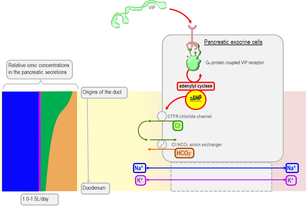
So alkaline are these secretions, that their loss (eg. by a pancreaticoduodenal fistula) leads to metabolic acidosis. Depending on whether you lean to Steward or to more classical interpretations of acid-base balance, this acidosis is either due to a loss of bicarbonate, or due to the reclamation of a large amount of chloride and the loss of sodium, leading to a decreased strong ion difference. This does have some relevance to the ICU trainee, as the uncontrolled leakage of pancreatic juice or duodenal contents is an important differential diagnosis of a normal anion gap metabolic acidosis, a popular topic for exam questions.
Water volume of pancreatic secretions
In total the pancreas is usually said to produce about 2500ml of fluid every day. If you collected this whole output in a jug, you'd be impressed by how clear and clean it looks. "Clear spring water" is how one article on pancreatic surgical complications has described it. It owes this appearance largely to being composed of mainly water, as apart from some dissolved enzymes there is usually nothing else in it, certainly nothing as garishly coloured as bile pigment. This water is thought to mainly come from duct cells, or at least that is what usually appears in textbooks, but acinar cells may play an equally important role.
The volume of pancreatic juice increases following a meal, and if there was no increase in the secretion of enzymes and bicarbonate, this postprandial juice would be weak and ineffective. What appears to happen instead is actually a disproportionate increase in the rate of enzyme production and chloride/bicarbonate exchange activity, such that the concentration of the active ingredients increases markedly with the rate of secretion (see the concentration/rate graphs above). The movement of water, and the increase in volume of the secretions, are therefore thought to be secondary phenomena, which occur in response to the increased concentration of duct lumen contents. Water moves from the acinar and ductal cells into the lumen along an osmotic gradient, following the movement of the electrolytes, and it probably does this through aquaporins (insofar as aquaporin-knockout mice seem to have some serious pancreatic insufficiency)
Corrosive properties of pancreatic secretions
From their pH, and from the fact that pancreatic enzymes are designed to attack and destroy organic molecules which until recently were plant and animal tissue, one might surmise that pancreatic secretions might have some pretty serious corrosive properties. How potent are they, a reasonable person might ask, and how effective would they be at dissolving a body?
The reader would be relieved to know that "no, nowhere and nobody" is the answer to the question "has anyone anywhere ever tried to regurgitate their pancreatic secretions on their victims as a forensic countermeasure". To be sure, pancreatic enzyme activation and "autodigestion" is blamed for the damage seen in pancreatitis, but overall these secretions seem to be relatively benign. Wickbom et al (1974) diverted pancreatic (well, duodenal) material in dogs, redirecting it to the ileum or stomach, and found the mucosae of these viscera was largely unchanged after four months of exposure. They even implanted the animal's spleens and kidneys into their gallbladder to see what would happen. After 58 days, these organs appeared to have made themselves at home in the biliary tree, being not only intact and functionally normal, but also having by that stage overgrown with normal-looking gallbladder mucosa. Exposure of the skin to pancreatic enzymes, such that might occur at the site of a pancreatic drain or fistula, does cause irritation and skin erosion, but over the course of days (Andersen et al, 1994). In short, this is not exactly xenomorph blood.
Digestive functions of pancreatic secretions
To put it simply, the pancreas is the main source of the enzymes which separate the molecules of the food you have eaten into something small enough to diffuse through a biological membrane. Ions in food don't need any help (they are small enough as it is), but amino acids fatty acids and sugars are inconveniently presented as huge unwieldy branching starch molecules, folded chunks of protein and insoluble globs of fat. Fortunately, the pancreas secretes a whole range of digestive enzymes which attack key points in these large molecules, disassembling them into small soluble fragments. There is no reason to remember these enzymes in any great detail; Normatov & Sentongo (2019) have this table, but it should be treated as an illustration rather than something to memorise:
.jpg)
(Henderson1, where it is adapted from, is not available anywhere. )
In addition to this digestive role, the alkaline fluid secreted by the pancreas is essential to neutralise the acidic secretions of the stomach. This reaction has several uses:
- Firstly, the fragile villi of the small intestine are too delicate to withstand the acerbic torments of your stomach acid, so something needs to neutralise it. When Hartzell et al (1962) ligated the pancreatic ducts of unsuspecting puppies, tthey found that duodenal pH would drop to somewhere between 1.0-2.0 for many hours following a meal. In pancreatectomized humans, this phenomenon produces "marginal ulceration" if the stomach content remains unbuffered, and prompts the necessary use of PPIs.
- Secondly, the function of all these abovementioned enzymes is optimal at a more neutral pH. In fact amylase and lipase are actually denatured at a pH below 4.5, which has implications for the replacement of enzymes in pancreatic insufficiency - if you just scorf a mug of pure pancreatic lipase, only 17% of it will still be active by the time it reaches the duodenum.
- Thirdly, the work of bile (micelle formation, etc) is dependent on pH, and does not work particularly well at a very acidic pH, as conjugated bile acids have a pKa of around 1-2, which means at the pH of stomach acid a fair proportion of their molecules would be in a useless non-ionised state.
.jpg)
Regulation of pancreatic exocrine function
The pancreas is not constantly leaking an uncontrolled stream of digestive secretions; rather, their production is carefully managed and coupled to the activity of the stomach, such that the release of pancreatic enzymes is proportional to, and coordinated with, gastric emptying.
Obviously this regulatory process has got to have some lead time built into it. This is not the gall bladder we are talking about, which can make a volume of bile immediately available by rapidly emptying. There is no reservoir of pancreatic juices to release, which means some time may pass between the regulatory signal and the appearance of pancreatic juice in the duodenum. Thus, in order for everything to have proper timing, some increase in the activity of the pancreas must take place as soon as food is contemplated, let alone eaten. Chandra & Liddle (2009) describe the following phasic response of the pancreas to the ingestion of a normal meal:
- Cephalic phase: 20-25% of the total pancreatic juice secretion occurs during this phase, and the main regulatory effector here is the vagus nerve. The juice secreted during this phase is apparently rich in enzymes but poor in bicarbonate, suggesting that the acinar cells are stimulated more than ductal cells.
- Gastric phase: 10% of the total pancreatic juice secretion occurs during this phase. It is again mainly the product of acinar cells, and appears to be mediated by the vagus nerve.
- Intestinal phase: the majority (60-80%) of pancreatic secretion occurs during this phase, and the main regulatory influences are neurohormonal. Specifically, secretin and cholecystokinin are the main stimulant hormones, and the vagovagal enterohepatic reflex
- Secretin release is stimulated by the arrival of acidic content into the duodenum, and it mainly stimulates the production of bicarbonate by the pancreatic duct cells
- Cholecystokinin release is mainly stimulated by intestinal lipid and protein content, and it stimulates the release of enzyme-rich acinar secretions, which makes logical sense.
- Vagovagal enteropancreatic reflex is an enteric reflex arc managed by the dorsal brainstem (Niebergall-Roth et al, 2006), and which is mainly stimulated by the direct activity of cholecystokinin on the pancreatic vagal nerve endings.
Pancreatic secretion is thought to be downregulated by sympathetic nervous system activity, though this is far from settled, and there are many conflicting data (for example, Love et al in 2007 listed studies that found noradrenaline increases, decreases, or completely fails to affect pancreatic secretion).
Somatostatin, and its analog octreotide, definitely decrease pancreatic exocrine secretions. Creutzfeldt et al (1986) found that the rate of enzyme synthesis can be suppressed by 80%. This has implications for the patient with the high-output pancreaticoduodenal fistula, where octreotide may be a valid management option.
References
Owyang, Chung, and John A. Williams. "Pancreatic secretion." Yamada's Textbook of Gastroenterology (2015): 450-473.
Mahadevan, Vishy. "Anatomy of the pancreas and spleen." Surgery (Oxford) 37.6 (2019): 297-301.
Cade, Jennifer E., and James Hanison. "The pancreas." Anaesthesia & Intensive Care Medicine 18.10 (2017): 527-531.
Epple, August, and Jack E. Brinn. "The Evolution of the Islet Organ." The Comparative Physiology of the Pancreatic Islets. Springer, Berlin, Heidelberg, 1987. 9-24.
Normatov, Inessa, and Timothy Sentongo. "Pancreatic malnutrition in children." Pediatric annals 48.11 (2019): e441-e447.
Lee, Min Goo, et al. "Molecular mechanism of pancreatic and salivary gland fluid and HCO3− secretion." Physiological reviews 92.1 (2012): 39-74.
Mangos, JOHN A., and NONA R. McSHERRY. "Micropuncture study of excretion of water and electrolytes by the pancreas." American Journal of Physiology-Legacy Content 221.2 (1971): 496-503.
HARTZELL, JOHN M., et al. "Changes in duodenal pH after exclusion of pancreatic secretions in dogs." Archives of Surgery 85.1 (1962): 142-151.
Butler, James R., et al. "Is antisecretory therapy after pancreatoduodenectomy necessary? Meta-analysis and contemporary practices of pancreatic surgeons." Journal of Gastrointestinal Surgery 19.4 (2015): 604-612.
Yamamoto, M., et al. "Water and enzyme secretion are tightly coupled in pancreatic secretion stimulated by food or CCK-58 but not by CCK-8." American Journal of Physiology-Gastrointestinal and Liver Physiology 288.5 (2005): G866-G879.
Gabbi, Chiara, et al. "Pancreatic exocrine insufficiency in LXRβ−/− mice is associated with a reduction in aquaporin-1 expression." Proceedings of the National Academy of Sciences 105.39 (2008): 15052-15057.
Wickbom, Gunnar, et al. "On the corrosive properties of bile and pancreatic juice on living tissue in dogs." Archives of Surgery 108.5 (1974): 680-684.
Singer, M. V., P. Layer, and H. Goebell. "Role of pancreatic enzymes in acute pancreatitis." Diagnostic Procedures in Pancreatic Disease. Springer, Berlin, Heidelberg, 1986. 67-74.
Andersen, P. H., et al. "Faecal enzymes: in vivo human skin irritation." Contact dermatitis 30.3 (1994): 152-158.
Love, Jeffrey A., Eunyoung Yi, and Tina G. Smith. "Autonomic pathways regulating pancreatic exocrine secretion." Autonomic Neuroscience 133.1 (2007): 19-34.
Creutzfeldt, Werner, et al. "Effect of somatostatin analogue (SMS 201-995, Sandostatin) on pancreatic secretion in humans." The American journal of medicine 82.5 (1987): 49-54.